39 muscle fiber model with labels
Muscle Models | Muscle Figures | Musculature Models - 3B Scientific Explore the muscles in 3D using the MICROanatomy™ Muscle Fiber. A single striated muscle fiber is magnified 10,000 times along with its neuromuscular end plate. Break down the muscular anatomy of the leg and hip (9-parts) and the muscles of the arm... 3/4 Life-Size Dual Sex Human Muscle Model on Metal Stand, 45-part - 3B Smart Anatomy $ 9,003.00 muscle labeling diagram muscle labeling diagram Muscles of Facial Expression Quiz we have 9 Pictures about Muscles of Facial Expression Quiz like Muscles of Facial Expression Quiz, Human Muscles Coloring Key | Educative Printable | Human body muscles and also Anatomy Games - Real Bodywork.
Muscles Labeling - The Biology Corner Typically, I would use straws and rubber bands to model fascicles and myofibrils. This year, students get a modified version of the lesson on Google Slides which explains how actin and myosin are organized and how those muscle fibers are grouped into bundles (fascicles) that make up a muscle. You are welcome to make a copy of these slides and modify them for you own class.
Muscle fiber model with labels
› en › trigger-point-therapyTrigger Point Therapy – That Is How We Treat Pain Jan 26, 2017 · a taut band (muscle fiber bundle) in the muscle; a pressure-sensitive area within the taut band; referred pain from a trigger point; a local twitch response of the trigger point or taut band in response to mechanical stimulation of the trigger point. These diagnostic criteria have been shown to have a high intertester reliability in trained ... en.wikipedia.org › wiki › Anabolic_steroidAnabolic steroid - Wikipedia Most steroid users are not athletes. In the United States, between 1 million and 3 million people (1% of the population) are thought to have used AAS. Studies in the United States have shown that AAS users tend to be mostly middle-class men with a median age of about 25 who are noncompetitive bodybuilders and non-athletes and use the drugs for cosmetic purposes. " Skeletal Muscle Fiber Labeling - Printable - PurposeGames.com This is a free printable worksheet in PDF format and holds a printable version of the quiz Skeletal Muscle Fiber Labeling. By printing out this quiz and taking it with pen and paper creates for a good variation to only playing it online.
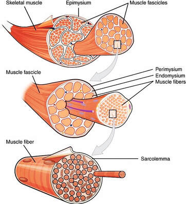
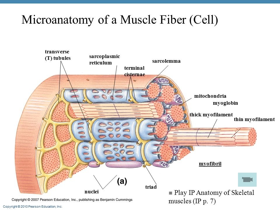
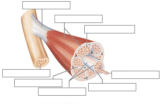
Muscle fiber model with labels. Structure of a Muscle Cell (Muscle Fibre) - ivyroses.com The nuclei of muscle fibres (' muscle cells ') are located at the edges of the diameter of the fibre, adjacent to the sarcolemma . As illustrated, a single muscle fibre may have many nuclei. Cytoplasm is present in all living cells. The cytoplasm present is muscle fibres ( muscle cells ) is called sarcoplasm. 9.2A: Skeletal Muscle Fibers - Medicine LibreTexts Skeletal Muscle Fiber Structure. Myocytes, sometimes called muscle fibers, form the bulk of muscle tissue. They are bound together by perimysium, a sheath of connective tissue, into bundles called fascicles, which are in turn bundled together to form muscle tissue. Myocytes contain numerous specialized cellular structures which facilitate their ... Myosin Orientation in a Muscle Fiber using Bifunctional Spin Labels ... Request PDF | On Feb 1, 2019, Yahor Savich and others published Myosin Orientation in a Muscle Fiber using Bifunctional Spin Labels with 4 Degrees Angular Resolution | Find, read and cite all the ... › articles › s43587/022/00250-8Characterization of cellular senescence in aging skeletal muscle Jul 15, 2022 · d, e, Representative images of quad muscle cross-sections stained for Laminin (d) and quantification of fiber size and distribution for young and old female mice (n = 4 per group) (e).
SAC A&P Model Key - Muscular System - austincc.edu Muscular System. M1 - Muscled Arm. M2 - Muscle Leg. M3 - Female Muscle Figure. M4 - Microanatomy Muscle Fiber. M5 - Muscle Figure. Skeletal Muscle Labeling MUSCULAR SYSTEM ANATOMY:Muscle Fiber With Neuromuscular Junction Model muscle junction neuromuscular fiber anatomy muscular system HLS [ Muscle Tissue, Skeletal Muscle, Ensheathment, Transverse muscle skeletal fascicles labeled histology tissue A Series Of Three Bones Showing The Basic Bone Markings. | Basic Muscles - Label - MUSTIB Flashcards | Quizlet Muscles - Label - MUSTIB STUDY Flashcards Learn Write Spell Test PLAY Match Gravity Created by ModelOrganism PLUS Animals Terms in this set (150) 1 - endomysium What is 1 pointing to? 2- skeletal muscle in cross-section What is the structure labeled 2? 3 - fasicle What is 3? 4 - perimysium What is 4? 5 - width of skeletal muscle fiber What is 5? Skeletal Muscle Fiber Model - Myofibrils - YouTube Skeletal Muscle Fiber Model - Myofibrils. 51,138 views Jan 7, 2009 This video was produced to help students of human anatomy at Modesto Junior College study our anatomical models.
Skeletal Muscle Histology Slide Identification and Labeled Diagram ... The skeletal muscle fibers are elongated, cylindrical and multinucleated cells whose length may vary in different animals. In this short guide, you will get a basic concept of skeletal muscle histology from the real slide and labeled diagram. You will also get the identification points of skeletal muscle histology slide with a little description here in this guide. Muscle Fiber Labeling Quiz - PurposeGames.com This is an online quiz called Muscle Fiber Labeling Quiz There is a printable worksheet available for download here so you can take the quiz with pen and paper. Your Skills & Rank Total Points 0 Get started! Today's Rank -- 0 Today 's Points One of us! Game Points 17 You need to get 100% to score the 17 points available Actions Add to Playlist › Muscle-Genuine-Protein-ShakeAmazon.com: Muscle Milk Genuine Protein Shake, Strawberries ... About this item . Contains twelve (12) 11 fl oz Cartons of Muscle Milk Genuine Protein Shakes. Packaging may vary. HELPS SATISFY HUNGER AND BUILD MUSCLE – Muscle Milk Genuine is an energizing protein shake that can be consumed as an on-the-go breakfast or anytime snack or to support post-workout recovery and muscle growth. Muscle Contraction & Sliding Filament Theory - TeachPE.com At a very basic level, each muscle fibre is made up of smaller fibres called myofibrils. These contain even smaller structures called actin and myosin filaments. These filaments slide in and out between each other to form a muscle contraction hence called the sliding filament theory! The diagram above shows part a myofibril called a sarcomere.
Blog
Muscle Fiber - an overview | ScienceDirect Topics The myofibrils are bundles of filaments arranged longitudinally parallel to the long axis of the muscle fiber. The various bands are named according to their position, appearance, or by how they rotate the plane of polarized light. Just as each muscle fiber contains many myofibrils, each myofibril is in turn composed of many filaments.

Pin on A&P
muscle fiber model labeling Diagram | Quizlet Start studying muscle fiber model labeling. Learn vocabulary, terms, and more with flashcards, games, and other study tools.

muscle fiber | Anatomy and physiology, Physiology, Health science
A spin label that binds to myosin heads in muscle fibers with its ... We have used an indane-dione spin label (2-[-oxyl-2,2,5,5-tetramethyl-3-pyrrolin-3-yl)methenyl]in dane-1,3-dione), designated InVSL, to study the orientation of myosin heads in bundles of chemically skinned rabbit psoas muscle fibers, with electron paramagnetic resonance (EPR) spectroscopy. After re …
Muscles - Runyan-Grigsby's Science Page
Learn all muscles with quizzes and labeled diagrams | Kenhub Human body muscle diagrams. Muscle diagrams are a great way to get an overview of all of the muscles within a body region. Studying these is an ideal first step before moving onto the more advanced practices of muscle labeling and quizzes. If you're looking for a speedy way to learn muscle anatomy, look no further than our anatomy crash courses .

What Did You Do Today at School?: Muscle Contraction Modeling
137,123 Muscle anatomy Images, Stock Photos & Vectors - Shutterstock Find Muscle anatomy stock images in HD and millions of other royalty-free stock photos, illustrations and vectors in the Shutterstock collection. Thousands of new, high-quality pictures added every day.

Pinterest • The world’s catalog of ideas
Skeletal Muscle Fiber Structure and Function - Open Textbooks for Hong Kong Figure 16.18 A skeletal muscle fiber is surrounded by a plasma membrane called the sarcolemma, with a cytoplasm called the sarcoplasm. A muscle fiber is composed of many fibrils packaged into orderly units. The orderly arrangement of the proteins in each unit, shown as red and blue lines, gives the cell its striated appearance.
U.jpg)
Anatomical Model, Muscle Fiber
Edible Muscle Fiber Model - Eclectic Homeschooling Edible Muscle Fiber Model Eclectic Homeschooling August 19, 2015 5th grade, our unit studies, science Sharing is caring! 141 As part of my son'smicrobiology study, he is learning about the different kinds of cells in the human body. He enjoys making things with food so we sat down to figure out how to make an edible muscle fiber model.

ANATOMY & PHYSIOLOGY I BIS 240: Muscle Fiber Model A
Muscular System Labeled Diagram Pictures, Images and Stock Photos Labeled Anatomy Chart of Male Triceps and Back Muscles on White... Labeled human anatomy diagram of man's arm, shoulder and upper back muscles in a posterior view on a white background. Cell potency. From Totipotent to Pluripotent, Multipotent, and... Development of fertilized egg and blastocyst to human fetus.

Neurolemmocyte On Skeletal Muscle Model - Human Anatomy - GUWS Medical
muscle diagram to label - anatomyclasspath.herokuapp.com muscle diagram to label The Echinoid Directory - Natural History Museum we have 9 Pics about The Echinoid Directory - Natural History Museum like Pin on Facial Anatomy, MUSCULAR SYSTEM ANATOMY:Muscle fiber with neuromuscular junction model and also Pin on Facial Anatomy.

1000+ images about A&P.2.Skin.Bone.Muscle on Pinterest | Models, Muscle and Anatomy
Labelled Diagram Of The Muscles In The Human Body - Anatomy Note Flexor carpi ulnaris is a fusiform muscle located in the anterior compartment of the forearm. It belongs to the superficial flexors of the forearm, along with pronator teres, palmaris longus, flexor digitorum superficialis and flexor carpi radialis. Flexor carpi ulnaris is the most medial of the superficial flexors. Flexor carpi radialis

BIO201 Muscle Tissue - Science 101 with Caston at Arizona State University - Tempe - StudyBlue
› Muscle-Milk-Protein-KnockoutAmazon.com: Muscle Milk Pro Advanced Nutrition Protein Shake ... About this item . Contains twelve (12) 11 Fl Oz cartons of Muscle Milk Pro Advanced Nutrition Protein Shakes. Packaging may vary. HELPS SATISFY HUNGER AND BUILD MUSCLE – The Muscle Milk Pro Advanced Nutrition Protein Shake is an energizing protein shake that can be consumed to support post-workout recovery and muscle growth and maintenance or to satisfy hunger in between meals.

Diagram on the left Setting the stage The events at the neuromuscular junction (NMJ) set the ...
Muscle Fibers: Anatomy, Function, and More - Healthline Each muscle fiber contains smaller units made up of repeating thick and thin filaments. This causes the muscle tissue to be striated, or have a striped appearance. Skeletal muscle fibers are...

Skeletal Muscle Fiber Model - Orientation - YouTube
Ultrastructure of Muscle - Skeletal - Sliding Filament - TeachMeAnatomy Ultrastructural Appearance of Skeletal Muscle. The striated appearance of skeletal muscle fibres is due to the organisation of two contractile proteins: actin (thin filament) and myosin (thick filament).. The functional unit of contraction in a skeletal muscle fibre is the sarcomere, which runs from Z line to Z line.A sarcomere is broken down into a number of sections:
muscle, circulatory, + nervous system Flashcards | Easy Notecards
Chapter 10 Muscle Tissue - Anatomy and Physiology Laboratory Manual for ... Skeletal muscle fibers can be identified by their multinucleate cytoplasm and striations (Figure 10.2) . Skeletal muscles move bones. Cardiac muscle fibers have one nucleus per cell, branched striations and intercalated disk (Figure 10.6). Cardiac muscles are found in the heart where they squeeze it to pump blood.

(Adopted from Atlas of Human Histology , by DiFiore, 3rd Ed., Lea & Febiger)
› ip › Muscle-Milk-Genuine-ProteinMuscle Milk Genuine Protein Powder, Vanilla Creme ... - Walmart Contains one (1) 1.93 pound canister (about 12 servings) of Muscle Milk Genuine Vanilla Creme Protein Powder. Packaging may vary. Shaker bottle sold separately. HELPS SATISFY HUNGER AND BUILD MUSCLE – Muscle Milk Genuine Protein Powder supplies your body with high quality protein to support post-workout recovery and muscle growth.

Do You Need To Lift Heavy To Build Muscle? | Training For Muscle vs Training For Strength
Skeletal Muscle Fiber - GetBodySmart Skeletal Muscle Fiber. Skeletal muscles are the types of muscle tissue that enable us with voluntary movements. They are attached to the bones of the skeleton by tendons. Skeletal muscle fibers have a striated (striped) appearance on histological sections because they are made up of smaller units called sarcomeres that run parallel to each other, giving the muscle the striated appearance.
Post a Comment for "39 muscle fiber model with labels"