43 ribosome diagram with labels
Bio 1113 - Unit 11 - Gene Expression Flashcards | Quizlet In the following diagram of a ribosome, assign the correct labels: Label 1: a tRNA attached to a polypeptide is found in this area of the ribosome Label 2: a tRNA attached to a single amino acid enters here Label 3: a tRNA that is not attached to anything exits here Label 4: a tRNA molecule Label 5: growing polypeptide Label 6: mRNA being ... › proteins › proteinProtein Targeting (With Diagram) | Molecular Biology ADVERTISEMENTS: Let us make an in-depth study of the protein targeting. After reading this article you will learn about: 1. Introduction to Protein Targeting 2. Signal Sequence 3. Transport of Proteins into ER 4. Signal Sequence Recognition Mechanism 5. Role of Golgi Complex in Protein Transportation 6. Transport of Proteins from Golgi to Lysosomes 7. […]
Ribosomes Vector Illustration. Anatomical and Medical ... Ribosomes vector illustration. Anatomical and medical labeled scheme. Explained closeup diagram.. Illustration about amino, educational, biogenesis, labeled, golgi, body - 122097933

Ribosome diagram with labels
Animal Cell Diagram with Label and Explanation: Cell ... Animal cell is a typical Eukaryotic cell enclosed by a plasma membrane containing nucleus and organelles which lack cell walls, unlike all other Eukaryotic cells. The typical cell ranges in size between 1-100 micrometers. The lack of cell walls enabled the animal cells to develop a greater diversity of cell types. Blood Histology Slides with Description and Labeled Diagram The blood is a specialized connective tissue that is fluid and circulates through the vascular channel. In the blood histology slide, you will find different types of cells with their specific features. This might be a short article where I will show you all the cells from the blood microscope slide with a labeled diagram and actual pictures. Plant Cell Diagram Ribosome Functions Ribosomes are a type of organelle. Ribosomes are small organelles of a cell having a dense feature and helps in protein fabrication. They are situated in the cytosol, some bound and free-floating to the membrane of the coarse endoplasmic reticulum. The ribosomes' structure is the same in all cells but smaller in prokaryotic cells.
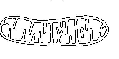
Ribosome diagram with labels. Structure of Ribosome (With Diagram) - Biology Discussion A bacterial ribosome is about 250 nm in diameter and consists of two subunits, one large and one small. Both subunits consist of one or more molecules of rRNA and an array of ribosomal proteins. ADVERTISEMENTS: Association of two subunits is called mono-some. The structure of prokaryotic ribosome is given in the figure 8.2 B. Structure of Ribosome - Biology Wise Diameter of Ribosome is 20nm. Their composition can be divided into two parts - 2/3 part of r-RNA (ribosomal RNA) and 1/3 part RNP (Ribosomal protein or Ribonuclep protein). Polypeptide chain is fabricated by translating mRNA (messenger RNA) with the aid amino acids that tRNA (transfer RNA) delivers. A Labelled Diagram Of Mitochondria with Detailed Explanation Mitochondria are a double-membrane-bound cell organelle found in most eukaryotic organisms. In all living cells, these cell organelles are found freely floating within the cytoplasm of the cell. The diagram of Mitochondria is useful for both Class 10 and 12. It is one among the few topics having the highest weightage of marks and is majorly ... Solved The ribosome in the diagram is in the process of ... The ribosome in the diagram is in the process of synthesizing a protein using directions transcribed from the DNA. Use the labels to identify each of the structures involved in translation and protein synthesis. Question: The ribosome in the diagram is in the process of synthesizing a protein using directions transcribed from the DNA.
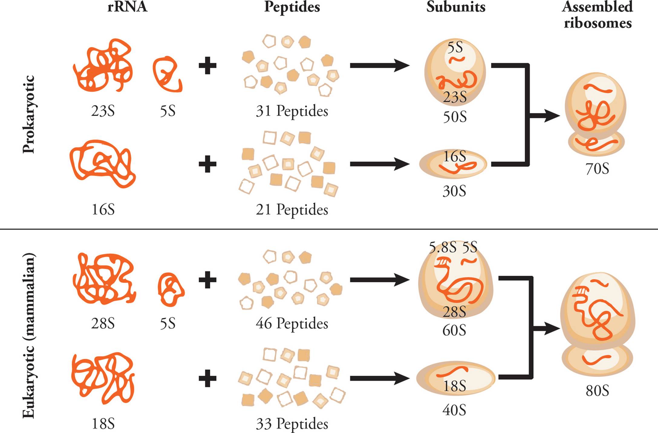
google-research.github.io › proteinferProteInfer: deep networks for protein functional inference We focus on Swiss-Prot to ensure that our models learn from human-curated labels, rather than labels generated by a computational annotation pipeline. Each protein in Swiss-Prot goes through a 6-stage process of sequence curation, sequence analysis, literature curation, family-based curation, evidence attribution, and quality assurance. quizlet.com › 394694271 › mastering-bio-6-flash-cardsMastering Bio #6 Flashcards | Quizlet Drag the appropriate tRNAs to the binding sites on the ribosome to show the configuration immediately before a new peptide bond forms. If no tRNA is bound to a site at that time, leave that binding site empty. b.) The diagram below shows an mRNA molecule that encodes a protein with 202 amino acids. Animal Cell Diagram | Science Trends An animal cell diagram is a great way to learn and understand the many functions of an animal cell. The diagram, like the one above, will include labels of the major parts of an animal cell including the cell membrane, nucleus, ribosomes, mitochondria, vesicles, and cytosol. Ribosomes: Structure, Composition, and Assembly (With Diagram) Ribosomes in the cytoplasm of eukaryotic cells have a sedimentation coefficient of about 80 S (MW about 4.5 x 10 6) and are composed of 40 S and 60 S subunits. In prokaryotic cells, ribosomes are typically about 70 S (MW about 2.7 x 10 6) and are formed from 30 S and 50 S subunits.
Label Transcription and Translation - Course Hero View Label Transcription and Translation - 7355418.pdf from BIOLOGY 101 at Harmony School of Innovation Fort Worth. 1. Label the diagram. DNA Protein Amino Acid Ribosome mRNA Codon tRNA Anticodon 2. PDF Quick Review Transcription and Translation - WPMU DEV label the diagram. 2. ... 910dnamrnait carries the genetic code from dna to ribosome to make a proteinit carries the amino acids to make proteinbecause the genetic code is the recipe to make a protein and is contained in a mrnacodons are in mrna and anti codons are groups of 3 bases in trnatranscription takes place in nucleus; translation takes ... molbiol-tools.ca › GenomicsGENOMICS - molbiol-tools.ca These multivariate analyses can be done using either taxonomic or automatically generated phenotypic labels and visualized using a variety of high quality graphical tools. The bacterial census data can be derived from 16S rRNA data, NextGen shotgun sequencing or even classical microbial culturing techniques. Includes a tutorial. protein synthesis diagram labeled - TheFitnessManual protein synthesis diagram labeled May 8, 2021 by Marie June Table of Contents 1. Transcription units the stage for Translation 2. Making protein is the aim of translation 3. Protein Chemistry Evaluation Quiz 4. Messenger RNA (mRNA) codes for proteins 5. Switch RNAs (tRNAs) deliver amino acids to the ribosome. - "protein synthesis diagram labeled"
Mitochondria - PurposeGames
Solved In the following diagram of a ribosome, assign the ... in the following diagram of a ribosome, assign the correct labels. 5' end of the mrna growing polypeptide a trna attached to a single amino acid ontors here large subunit atrna attached to a polypeptide is found in this area of the nibosome a trna that is not attached to anything exits hore 3' end of the mrna a trna moleculo mossenger rna being …
The Anatomy and Physiology of Animals/The Cell Worksheet - WikiEducator
Protein Synthesis Labeling.pdf - 1. Label the diagram ... Protein DNA Amino Acid mRNA Codon tRNA Ribosome Anticodon 2. Label the diagram. Protein Amino Acid Large subunit - rRNA tRNA mRNA codon small subunit rRNA 3. What is the role of mRNA in the process of protein synthesis? The role of mRNA in protein synthesis is that it carries copies of genetic instructions (that tell the cell how to assemble ...
Molecular Biology - MCAT Biology and Biochemistry
Cell Organelles- Definition, Structure, Functions, Diagram In the case of prokaryotic cells, the ribosomes are of the 70S with the larger subunit of 50S and the smaller one of 30S. Eukaryotic cells have 80S ribosomes with 60S larger subunit and 40S smaller subunit. Ribosomes are short-lived as after the protein synthesis, the subunits split up and can be either reused or remain broken up.

Copy of "Nucleus and Transcription: The RNA polymerase b..."
Labeled Plant Cell With Diagrams - Science Trends The ribosomes are created in the nucleolus of the cell. Ribosomes are made out of two smaller subunits, a large ribosomes subunit and a small ribosomal subunits. The transfer RNA or tRNA encodes the correct series of genetic instructions into the mRNA or messenger RNA, which is what ensures that the right proteins are created.

Drag The Labels Onto The Diagram To Identify The Stages Of The Cell Cycle. — UNTPIKAPPS
Ribosome - Wikipedia Prokaryotic ribosomes are around 20 nm (200 Å) in diameter and are composed of 65% rRNA and 35% ribosomal proteins. Eukaryotic ribosomes are between 25 and 30 nm (250-300 Å) in diameter with an rRNA-to-protein ratio that is close to 1.
Post a Comment for "43 ribosome diagram with labels"